2025 Larsson, M. & D. Falk. Direct effects of bipedalism on early hominin fetuses stimulated later musical and linguistic evolution, Current Anthropology, forum article with international commentaries, in progress.
2024 Falk, D. & A. Marom. The DNH7 endocast of Paranthropus robustus from Drimolen, South Africa: Reconsidering the functional significance of an enlarged occipital-marginal (O/M) sinus system in robust australopithecines. American Journal of Biological Anthropology: e25010. https://onlinelibrary.wiley.com/doi/pdf/10.1002/ajpa.25010
2023 Nicole Labra, Yann Leprince, Denis Rivière, Mathieu Santin, Jean François Mangin, Lou Albessard-Ball, Amélie Beaudet, Emiliano Bruner, Douglas Broadfield, Kristian Carlson, Zachary Cofran, Dean Falk, Emmanuel Gilissen, Aida Gomez Robles, Aurélien Mounier, Simon Neubauer, Alannah Pearson, Carolin Röding, Yameng Zhang, Antoine Balzeau. What do brain endocasts tell us? A comparative analysis of the accuracy of sulcal identification by experts and perspectives in palaeoanthropology. Journal of Anatomy, 00, 1-23: https://doi.org/10.1111/joa.13966
2020 Gunz, P., S. Neubauer, D. Falk, P. Tafforeau, A. Le Cabec, T.M. Smith, W.H. Kimbel, F. Spoor, Z. Alemseged. Australopithecus afarensis endocasts suggest ape-like brain organization and prolonged brain growth. Science Advances 6 (14), April 1.
2018 Falk, D., C.P.E. Zollikofer, M. Ponce de Leon, K. Semendeferi, J.L. Alatorre Warren, W.D. Hopkins. Identification of in vivo Sulci on the external surface of eight adult chimpanzee brains: Implications for interpreting early hominin endocasts. Brain, Behavior and Evolution: https://www.karger.com/Article/FullText/487248
2016 DeanFalk Evolution of brain and culture: The neurological and cognitive journey from Asutralopithecus to Albert Einstein, Journal of Anthropological Sciences
2014 Dean Falk. Interpreting sulci on hominin endocasts: old hypotheses and new findings. Frontiers in Human Neuroscience
Hominin paleoneurologists are scientists who investigate brain evolution by studying the fossil record of early human ancestors. How, you might wonder, can one learn about the evolution of the cognitive traits that define humans (language, for example) from fossils?
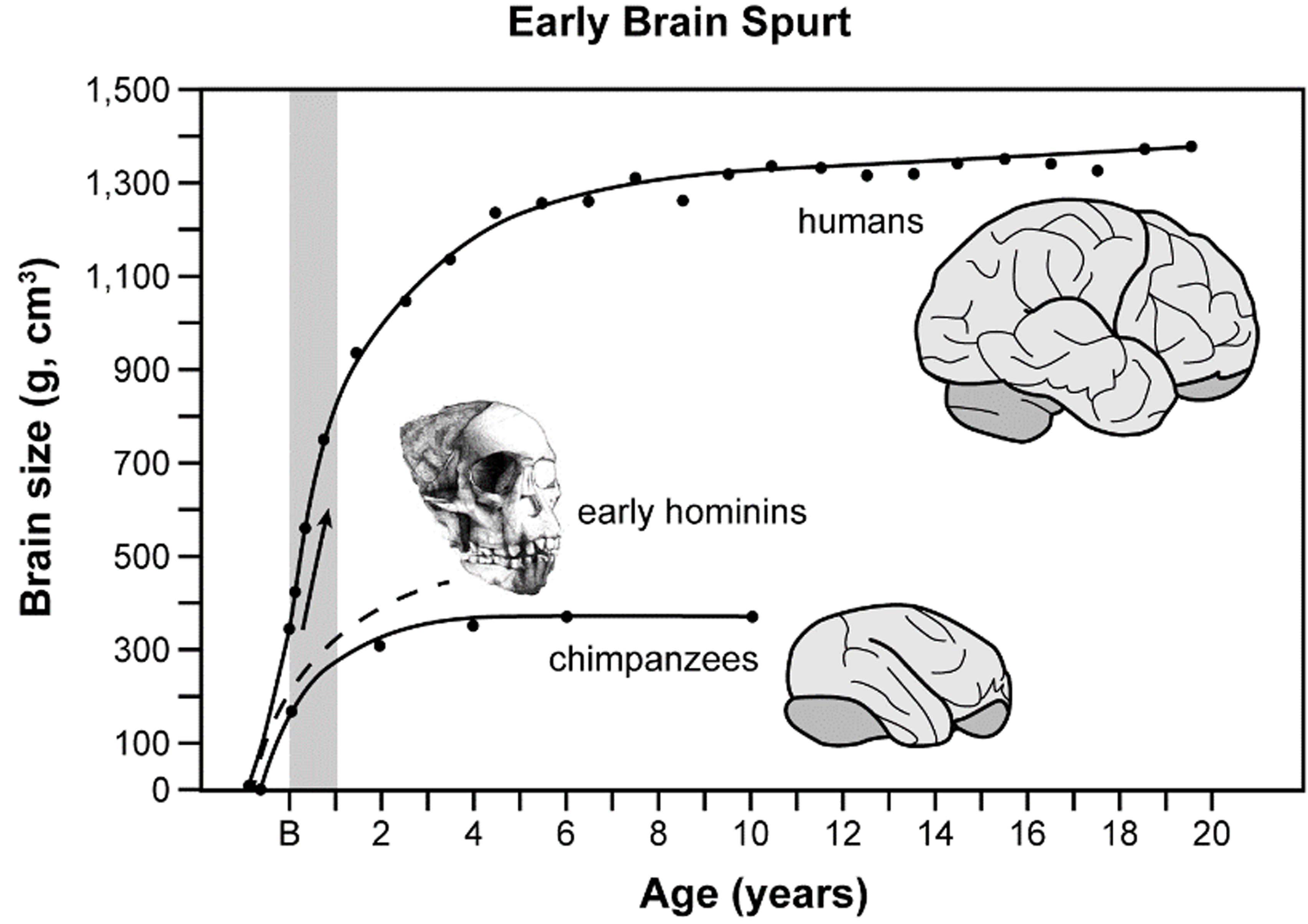
Human ancestors evolved larger brains during approximately the past three million years as growth rates in newborns’ brains (“brain spurts,” shaded column) became steeper over time (arrow). B, birth; Age, in years for each individual.
This kind of research requires interpretation of details that are imprinted in stone, which are often murky and difficult to decipher. Partly for this reason, paleoneurologists argue passionately about how, when, and why human brains evolved to their present-day state.
The evidence that paleoneurologists interpret comes mainly from the fossilized braincases of our early ancestors. Traditionally, the volume of the braincase (its cranial capacity in cubic centimeters, cm3) has been used as a surrogate for brain size. This practice remains widespread even though the cranial capacity of a skull is somewhat of an overestimate for brain size because braincases contain fluids and membranes in addition to brains. Nevertheless, cranial capacities are easy to measure and provide an excellent window into the evolution of brain size when they are plotted against time on a graph. From such data, we know that brain size increased over many millions of years in a wide variety of mammals, including those from major lineages in our own group, the primates.
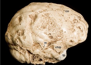
A nearly complete endocast that provides information about the right side of the cerebral cortex of an early human relative (Australopithecus africanus).
Compared to most other animals, humans have very large brains in absolute terms (by far, the largest of any primate) and also with respect to the sizes of their bodies (called relative brain size, RBS). Plots of cranial capacities over time reveal that average brain size in living people (around 1350-1400 cm3) is three to four times that of our early ancestors (called australopithecines) who lived in Africa over three million years ago. As our ancestors evolved, natural selection favored an exceptional upward trend in absolute brain size. For a variety of reasons, many scientists view absolute brain size (as opposed to RBS) as the single best measure for tracking the evolution of cognition in our early ancestors. At the moment, it appears that brain size may have topped out in our species, and even declined in some modern groups compared to our late relatives, the Neanderthals. As with computers, size is not everything when it comes to brains—the quality and combination of the brain’s various regions, neurochemicals, and connections (neurological organization) is also extremely important.
Some of the most interesting information about the brains of our ancestors comes from internal molds of braincases, known as endocranial casts or endocasts. An endocast reproduces the shape and, with luck, some of the details of the external surface of the brain (the cerebral cortex) that were imprinted on the walls of the braincase when the individual was alive. This is fortunate for paleoneurologists because the human cerebral cortex is the part of the brain that facilitates highly evolved functions such as language and rational problem solving. For this reason, paleoneurologists spend a good deal of time scrutinizing endocasts from early human relatives, some of whom lived from five to seven million years ago!
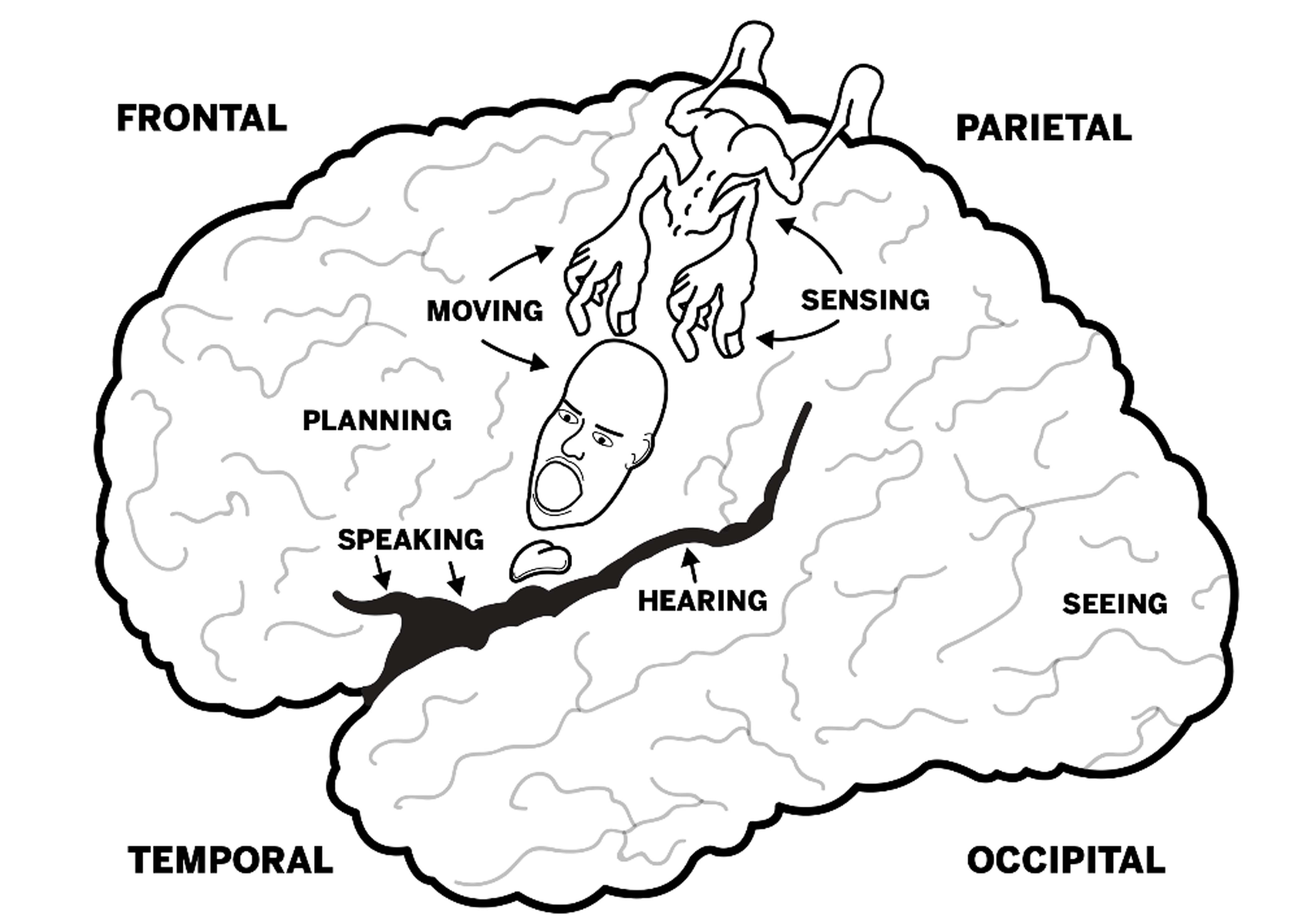
Lobes of the brain and locations for some basic functions (left side of human brain). The little person (homunculus) illustrates the locations that facilitate movement and reception of sensory information for specific body parts (mostly on the opposite side of the body).
Viewed from the outside, brains are composed of different lobes that (broadly speaking) facilitate different basic functions. For example, vision is processed in the occipital lobes at the back of the brain. Brains also have right-left differences, and these are particularly marked in humans—both visually (brains appear lopsided when viewed from the top) and functionally (in the majority of people, language is processed mostly by the left side of the brain, while music depends more on the right side). The regions between the primary cortices that represent the basic functions such as hearing, seeing, and so on are called association cortices. Association cortices synthesize and analyze information they receive from other parts of the brain (including primary cortices), and are thought to have been of paramount importance for the evolution of advanced cognition in humans.
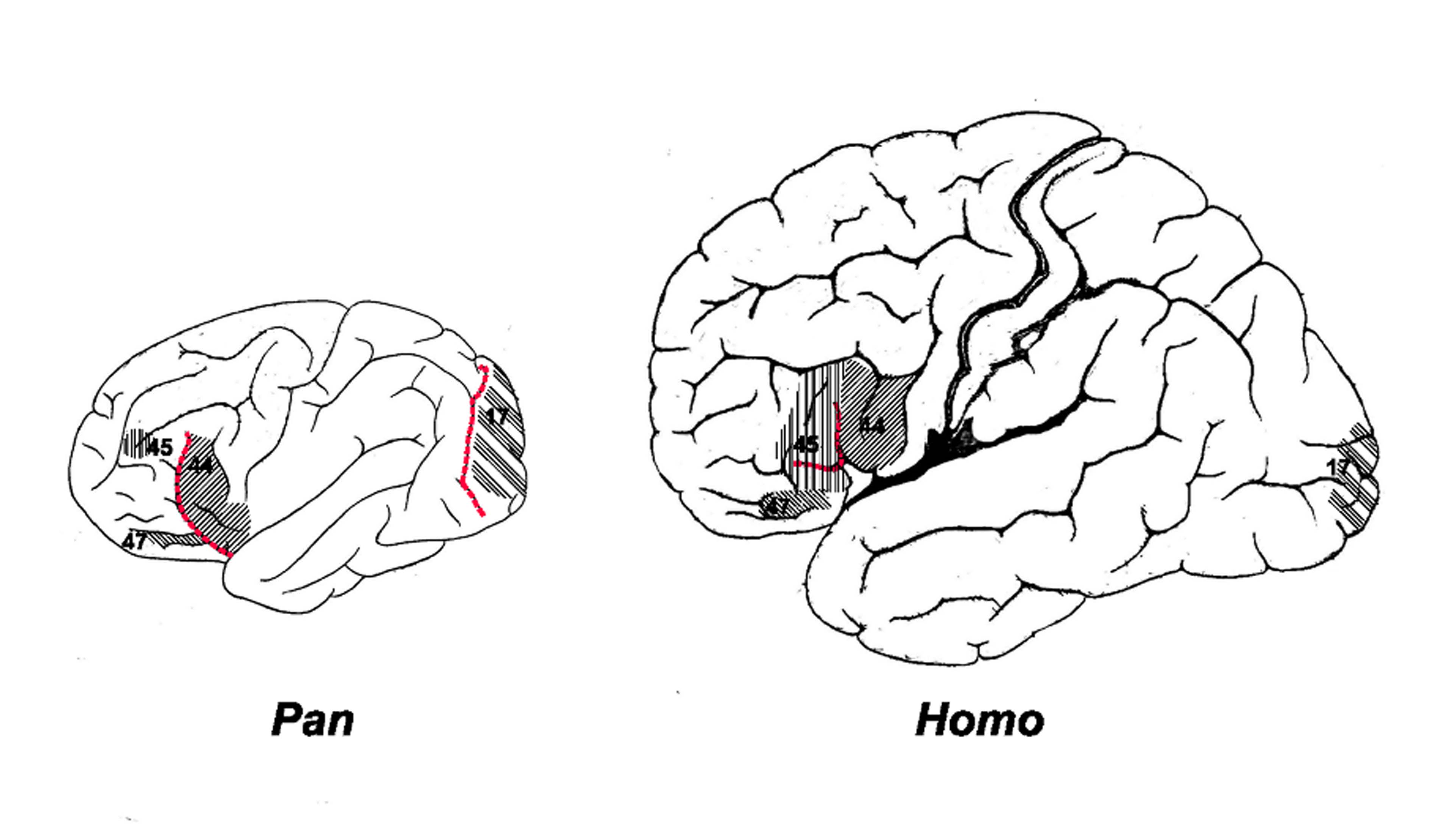
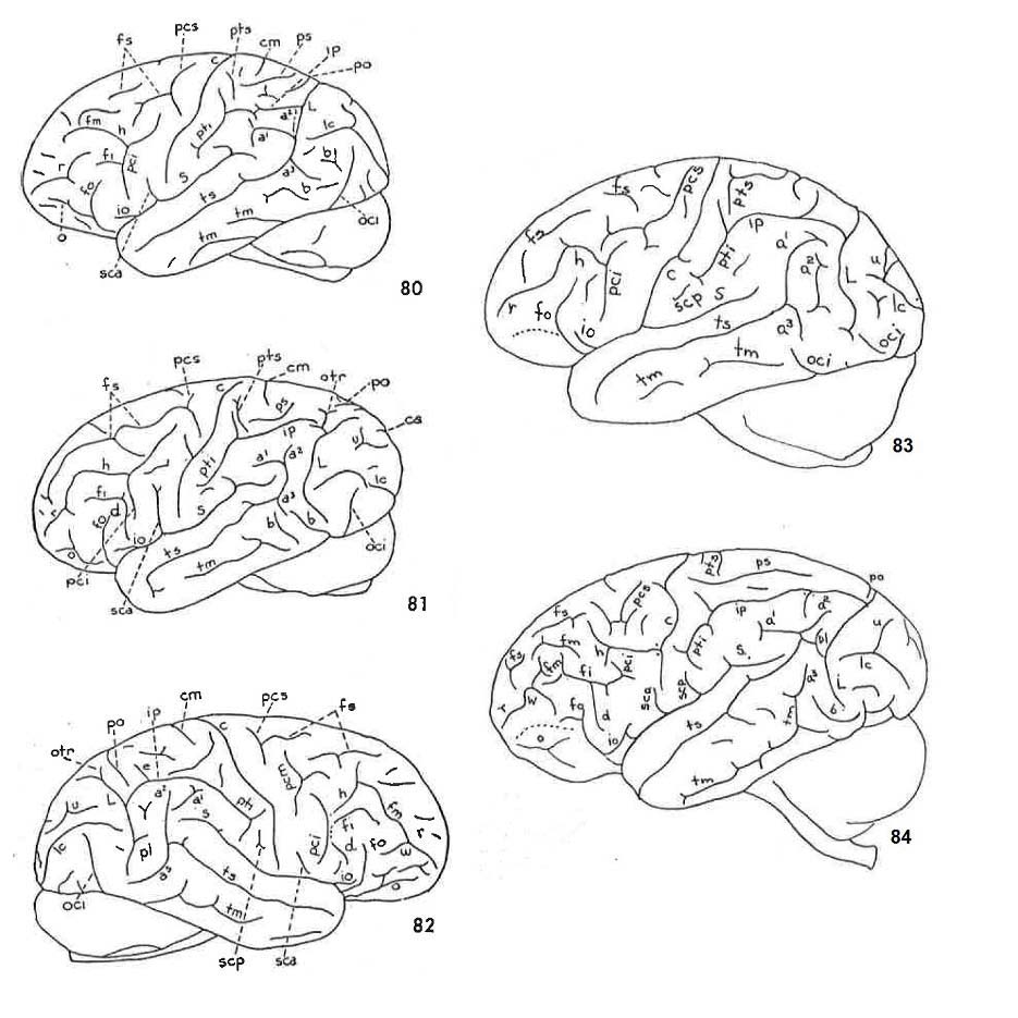 The cerebral cortex of most primates (and, indeed, most mammals) consists of convolutions of gray matter (gyri) that are separated by grooves (sulci). The patterns of sulci (sulcal patterns) can be very telling and, for this reason, paleoneurologists are keenly interested in the traces of sulci that are sometimes reproduced on endocasts from the skulls of our early ancestors. Genetically speaking, chimpanzees (Pan) are the closest living “cousins” of people (Homo). It is, therefore, interesting to compare the sulcal patterns that are typical for the two groups to get an idea of the changes that must have occurred at some point in the cerebral cortices of our ancestors. The line drawings shown here illustrate sulcal patterns for five chimpanzees that were discussed in John Connolly’s splendid 1950 monograph, External Morphology of the Primate Brain.
Unfortunately, the patterns of the main (named) sulci of humans do not differ very much from those of chimpanzees and the other great apes. In fact, there are only two places in the brain where the typical sulcal pattern of apes and humans differ significantly. (1) The frontal lobes of apes have large curved (fronto-orbital) sulci in front of Brodmann’s area (BA) 44 that are lacking in human brains, which instead have two little sulci that form two sides of a triangular patch of cortex (BA 45). On the left side of the human brain, this patch is part of Broca’s speech area. (2) Apes and people also differ in the back parts of their brains. In apes, a large crescent-shaped sulcus (the Affenspalte or lunate sulcus) courses just in front of the primary visual cortex (BA 17). In humans, BA 17 has been displaced toward the back of the brain. As this reorganization evolved, the Affenspalte was lost. A comparison of ape and human sulcal patterns suggests that, as the brains of our ancestors evolved and became larger, the cerebral cortex at both ends of the brain expanded and became reorganized in ways that, presumably, facilitated hominins’ emerging cognitive abilities (such as language). The trick, of course, is to try to detect when, exactly, these changes in cortical sulcal patterns occurred. So far, we do not know—partly because the relevant sulci do not happen to reproduce well on endocasts.
In sum, what paleoneurologists can potentially learn about human brain evolution from fossils is confined to information about (1) the evolution of brain size and (2) how and when limited parts of the cerebral cortex became reorganized. As noted, the cerebral cortex is a highly evolved part of the human brain. Among other things, it facilitates conscious thought, planning, language, social skills, and scientific, artistic, and musical creativity. Because it is the most superficial part of the brain, the cerebral cortex may leave imprints in skulls, which are sometimes reproduced on endocasts. For these two reasons, paleoneurologists tend to be cortical chauvinists. Consequently, the internal structures of the brain have not received as much attention from paleoneurologists. These parts of the brain also evolved, and they are extremely important for processing memories, gut-level feelings, and social interactions in ways that set humans apart from other animals.

